| Watermelon Diseases | ||||||||||||||||||||||||||||||||||||||||||||||||||
|---|---|---|---|---|---|---|---|---|---|---|---|---|---|---|---|---|---|---|---|---|---|---|---|---|---|---|---|---|---|---|---|---|---|---|---|---|---|---|---|---|---|---|---|---|---|---|---|---|---|---|
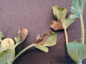
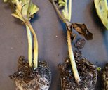
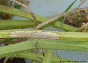
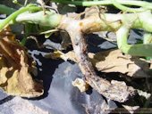
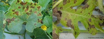
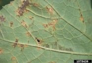
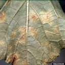
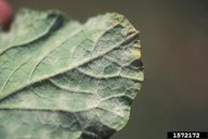
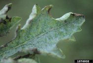
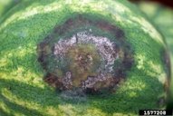
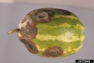
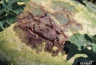
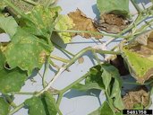
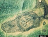
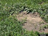
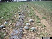
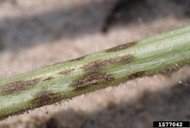
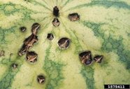
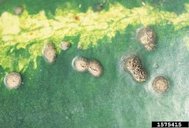
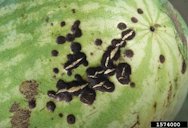
| Back to Watermelon Page 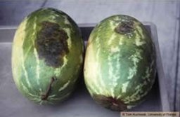 Fig. 1 Black rot symptoms on watermelon, honeydew melon, and butternut squash 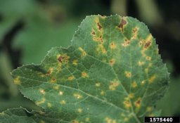 Fig. 7  Sporulation of Pseudoperonospora cubensis on underside of pumpking (cv. Howden) leaf 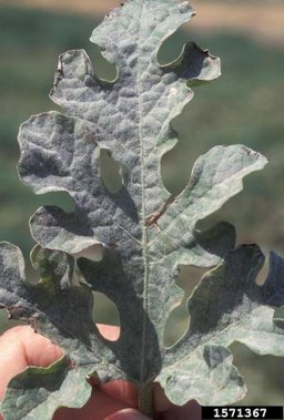 Fig. 10  Watermelon leaf showing an even distribution of powdery mildew (Podosphaera xanthii) over the entire leaf surface 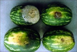 Fig. 14  Various stages of fruit rot of watermelon caused by Phytophthora capsici. Bottom, early symptoms; top, advanced symptoms. 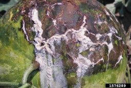 Fig. 18 Mature fruit showing lesion, cracking, foaming bacterial ooze and copper spray residue on fruit surface. Bacterial ooze has been colonized by the fungus Geotrichum candidum. 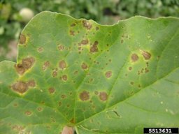 Fig. 22  Alternaria leaf blight, many foliar lesions of varying ages 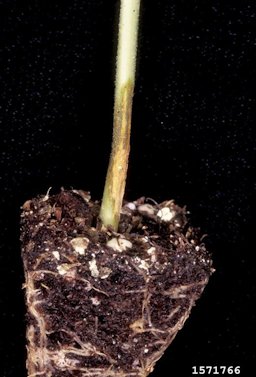 Fig. 24  Watermelon transplant showing a water-soaked lesion on the lower stem. Symptoms are consistent with infection by Rhizoctonia solani. 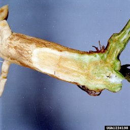 Fig. 25 Fusarium wilt symptoms on watermelon stem 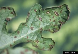 Fig. 27  Note the cracking of necrotic tissue, irregular lesion margins and shot hole effect
|
The
most important fungal diseases are gummy stem blight (caused by Didymella bryoniae/Phoma
cucurbitacearum) and downy mildew (caused by Pseudoperonospora cubensis).
Some consider gummy stem blight to be the most troublesome disease on
watermelons in Florida. Other occasional or minor diseases include
phytophthora blight (caused by Phytophthora
capsici), bacterial fruit blotch (caused by Acidovorax avenae
subsp. citrulli),
alternaria leafspot (caused by Alternaria
cucumerina), seedling blight (caused by Pythium spp., Rhizoctonia solani,
and Fusarium
spp.), fusarium wilt (caused by Fusarium
oxysporum f.sp. niveum), angular leafspot (caused by Pseudomonas syringae),
anthracnose (caused by Colletotrichum
orbiculare), rind necrosis (usually caused by Erwinia spp.), and
powdery mildew (caused by Erysiphe
cichoracearum).
Blossom end rot, a physiological disorder related to calcium deficiency
and water stress, is also an occasional problem. Finally, Cercospora leafspot
(caused by Cercospora
citrullina) and southern blight (also called southern stem
rot or white mold and caused by Sclerotium
rolfsii) are only rarely seen on watermelons in Florida
(Hochmuth et al. 1997; Maynard 2003; Roberts and Kucharek 2005). 1 Gummy Stem Blight (Fig. 1) Caused by Didymella bryoniae/Phoma cucurbitacearum Gummy stem blight (caused by D. bryoniae/P. cucurbitacearum) is one of the most important diseases of watermelons in Florida. It can affect most aboveground parts of the watermelon plant. Symptoms may be difficult to distinguish from other foliar diseases and include brown to black leaf spots, stem cankers, or fruit spots. Lesions eventually develop black spots, which are the fruiting bodies of the fungus (pycnidia). On watermelon, the disease is referred to as black rot. It produces water-soaked spots on the fruit, which leak gummy material (Maynard and Hopkins 1999; Newark et al. 2014; Roberts and Kuchaek 2005). 1
Fig. 2. Necrosis on cantaloupe transplants due to gummy stem blight Fig. 3. Water-soaked regions on the stem of cantaloupe transplants due to gummy stem blight Fig. 4. Canker on watermelon stem due to gummy stem blight Fig. 5. Gummy red exudation from a D. bryoniae infected cantaloupe stem Fig. 6. Brown, irregularly shaped lesions containing a concentric ring pattern can be seen on cantaloupe and watermelon leaf margins and interveinal areas with high moisture retention Further Reading Management of Gummy Stem Blight (Black Rot) on Cucurbits in Florida, University of Florida pdf Downy Mildew (Fig. 7) Caused by Pseudoperonospora cubensis Downy mildew (caused by Pseudoperonospora cubensis) is another key fungal disease of watermelons in Florida. It occurs principally in South Florida, and its likelihood of occurrence in the state decreases with distance to the north. It occurs every year in South Florida, while in North Florida its presence is sporadic. Ideal conditions for the development of downy mildew include nighttime temperatures of 55 °F–75 °F (12.8 °C–23.9 °C) and relative humidity above 90%. It is possible for watermelons planted in the fall and spring may be infected with downy mildew as early as the appearance of the first true leaves (Newark et al. 2016; Roberts and Kucharek 2005). 1
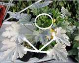
Fig. 8. Sporulation of P. cubensis on underside of pumpkin (cv. Howden) leaf Fig. 9. Downy mildew on melon leaf Powdery Mildew (Fig. 10) Caused by Podosphaera xnathii or Erysiphe cichoracearum Powdery mildew (caused by P. xnathii or E. cichoracearum) is a common and serious disease of watermelons in Florida. Since no resistant cultivars are available for this disease, fungicides are the primary means for management. Typical symptoms are the production of white, powder-like colonies on the leaf surface which are found on the leaves and petioles. Yellow spots can also be noticed the leaf, which can be confused with other diseases, such as downy mildew or bacterial leaf spots. In severe cases, the fungus will colonize the whole leaf, leading to death. 1 Dry conditions, usually followed by periods of free moisture at night, tend to promote the reproduction and spread of this pathogen. Spores are produced in chains on the leaf surface and are removed from the leaf by wind. These spores then infect watermelon leaves and the cycle repeats multiple times throughout the season. The pathogen can develop and spread quickly under favorable conditions with symptoms appearing three days after infection. 1
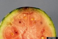
Fig. 11. Squash plant infected with powdery mildew; notice how old leaves are completely covered with talc-white powdery mildew (arrows), whereas new leaves appear to be free of this disease (circle) Management of Cucurbit Downy Mildew in Florida, University of Florida pdf Phytophthora blight (Fig. 14) Caused by Phytophthora capsici Phytophthora blight (caused by P. capsici) occurs sporadically in Florida, but the disease can spread rapidly if the weather is favorable to the fungus, causing serious losses. In 1998, the disease was widespread and severe on several vegetable crops in Florida. In the southwest region of the state (Lee, Collier, and Hendry Counties), 25% of watermelon plants in some surveyed fields were found to have the disease, and disease incidence on watermelons in Manatee County was 36%. Previously, phytophthora blight had affected only the fruit of watermelons, but during the 1998 outbreak, many watermelon plants were affected and died in Manatee County, regardless of plant age. 1 Watermelon foliage is less susceptible to P. capsici than that of summer squash. However, all stages of watermelon fruit are highly susceptible. Early symptoms of fruit rot include rapidly expanding, irregular, brown lesions which become round to oval. Concentric rings may occur within a lesion. The centers of rotted areas are covered with grayish fungal-like growth, while the outer margins of lesions appear brown and water-soaked. The entire fruit eventually decays. Initial symptoms of bacterial fruit blotch of watermelon are similar to those caused by P. capsici. However, after lesions expand, the two diseases can be easily separated because of the presence of extensive rind cracking, absence of fungal-like growth, and other symptoms associated with bacterial fruit blotch. Phytophthora rot is characterized by abundant fungal-like growth accompanied by little or no cracking. 2
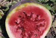
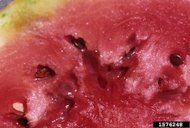
Fig. 15. Cross-section through watermelon fruit showing the depth and breadth of a Phytophthora blight lesion Fig. 16. Fruit lesion Fig. 17. Fruit with several large legions Further Reading Vegetable Diseases Caused by Phytophthora capsici in Florida, University of Florida pdf Bacterial Fruit Blotch (Fig. 18) Caused by Acidovorax avenae subsp. citrulli Bacterial fruit blotch (caused by A. avenae subsp. citrulli) occurs sporadically, affecting only a limited number of fields, but it can cause severe losses where it occurs. Also called greasy fruit spot or watermelon fruit blotch, this bacterial disease is a relatively new problem in Florida watermelon production. The pathogen produces both a leafspot and a fruit spot, and symptoms can occur on seedlings, leaves, and fruit. When it attacks seedlings, water-soaked lesions form, and the seedling can collapse and die. Found principally along the major veins, lesions on the leaves are light to reddish brown and can occur throughout the season. Although they are generally inconspicuous and do not usually contribute to defoliation, leaf lesions serve as reservoirs of the bacteria that later infect the fruit. On the watermelon fruit, lesions start as very small, water-soaked areas that usually do not appear until close to harvest. Later, they enlarge, and within two weeks, they may cover the entire upper surface of the fruit. Cracks may later appear on the rind, which may show internal discoloration, and the whole fruit may rot within, as secondary pathogens enter the open lesions (Roberts and Kucharek 2005; Somodi et al. 1991; Hopkins, Cucuzza, and Watterson 1996; Hopkins 1991; Frankle, Hopkins, and Stall 1993). 1
Fig. 19. Mature fruit showing lesion, deep cracking, bacterial ooze and copper spray residue on fruit surface Fig. 20,21. Cross section through mature fruit shows external and internal damage Alternaria Leafspot (Fig. 22) Caused by Alternaria cucumerina Alternaria leaf spot (caused by A. cucumerina) is a minor disease on Florida watermelons. Symptoms begin on the upper surface of older leaves as very small yellow or tan spots that may be surrounded by light green or yellow halos or by a water-soaked area. The spots later grow up to 3/4 of an inch (2 cm) in diameter and turn brown in color. Similar in appearance to gummy stem blight, the lesions are the source of spores spread primarily by the wind. Under severe infestations, the disease produces leaf curling, defoliation (which leaves the fruit susceptible to sunscald), and premature ripening. Lower yields, lower fruit sugar, and fruit deformity may occur (Roberts and Kucharek 2005). 1
Fig. 23. Alterneria leaf blight of cantaloupe Fig. 24. On cucumber Angular Leaf Spot Caused by Pseudomonas syringae pv. lachrymans/Pseudomnas syringae Symptoms are found on the leaves, stems, and fruit. Spots on the leaves are typically angular and water- soaked. Free moisture allows the bacteria to ooze from the spots, which may, upon drying, leave a white residue. These spots of dead tissue will occasionally drop away from the healthy tissue leaving irregular ‘shotholes’ in the leaves. 3 The spots on the fruit are generally smaller, nearly circular and slightly depressed. Early external fruit infections may be so small as to be impossible to cull out during packing. Internal symptoms, however, are quite obvious. The internal flesh discolors (brown) from below the skin lesion down to the seed layer within the fruit and may run the entire length of the fruit. Older fruit lesions turn white with an obvious tissue cracking. This disease is apt to be severe during wet springs and is rain-splash dispersed. The causal bacterium can survive in infected crop debris. The bacterium is seedborne. 3 Seedling Blight (Fig. 24) Caused by Pythium spp., Rhizoctonia solani, and Fusarium spp. Seedling blight (caused by Pythium spp., Rhizoctonia solani, and Fusarium spp.) can kill seedlings before or after they emerge. Rot symptoms, either wet or dry, are observed in the presence of this disease. The lesions resulting from seedling blight caused by Rhizoctonia solani (Fig. 23) are reddish-brown to orange and appear sunken. Pythium spp. cause shoots or roots to appear gray and water soaked. The incidence of seedling blight is higher when watermelons are planted in cool soils because seedlings that emerge slowly are more susceptible to pathogenic fungi (Roberts and Kucharek 2005). 1 Fusarium Wilt (Fig. 25) Caused by Fusarium oxysporum f. sp. niveum The causal fungus is soilborne and may infect the cucumber at any stage of growth. Freshly seeded cucumbers may damp-off below ground or as newly emerged seedlings. If older plants are infected prior to vine elongation, the entire plant will exhibit a mid-day wilt. After several days of wilting, leaves will tip burn followed closely by a complete wilt and plant death. Plants infected in the vining stage of growth will often wilt in only one or two runners. Diagnostic field symptoms are the wilting syndrome and the brown discoloration of the vascular tissue within the lower stem when split open. In moist weather, the whitish- pink fungal mycelium may be observed on the outside of the lower stems. 3
Fig. 26. Fusarium wilt symptoms in a watermelon field Fig. 27. Symptoms of fusarium wilt in a watermelon field Further Reading Race 3, a New and Highly Virulent Race of Fusarium oxysporum f. sp. niveum Causing Fusarium Wilt in Watermelon, Texas A&M University System, University of Maryland, University of Delaware and the US Department of Agricultural Research Service pdf Anthracnose Caused by Colletotrichum orbiculare In the past, anthracnose (caused by C. orbiculare) was a serious disease in Florida watermelon production, but the use of resistant varieties has limited its impact. Three races of anthracnose are known (races 1, 2, and 3), and some varieties show resistance to some races but not others. Anthracnose race 2 has caused serious damage to watermelons in the southeastern United States. When it does occur, anthracnose can destroy the entire field if not controlled, particularly after several days of warm, rainy weather (Bertelsen et al. 1994; Roberts and Kucharek 2005). All above ground plant parts may be affected by this fungal disease, and infected plants may die under severe conditions. Early symptoms of the disease include angular, brown to black leaf spots on older leaves, similar in appearance to those of gummy stem blight or downy mildew. Tan, oval-shaped lesions may appear on the stems. Spores from leaf and stem lesions later infect the fruit, producing sunken, water-soaked spots. 1 Disease development is greatest during humid, rainy weather. Spores are spread by wind, splashing rain, people, and machinery. The fungus, which can be seedborne, survives between crops on infected plant debris and volunteer plants (Bertelsen et al. 1994; Roberts and Kucharek 2005; Maynard and Hopkins 1999). 1
Fig. 28. Anthracnose on watermelon stems. Note the black stromata in the lesion at the center of the frame Fig. 29, 30. Cracking of lesion is characteristic of anthracnose Fig. 31. Fruit lesions Further Reading Anthracnose on Cucurbits in Florida, University of Florida pdf Damping-Off Pythium spp., Fusarium spp., Rhizoctonia spp. Seed fails to germinate due to rapid colonization of seed by soilborne fungi and fungal-like organisms. Excavated seed will be rotted and soft, often with evidence of fungal mycelium. Young, newly emerged seedlings often collapse at soil line and topple over. The stems may exhibit an obvious discoloration ranging in color from a reddish- brown to black and may be dry or mushy to the touch depending on the soil fungus involved. 3 Belly Rot Caused by Rhizoctonia solani This disease is caused by a common soilborne fungus which infects the ground (belly) side of fruit. Young cucumbers exhibit a yellow reddish-brown, superficial discoloration which develops into a sunken, irregular lesion or pit in the fruit underside. Mature fruits develop a large, water-soaked decay. This disease proceeds rapidly above 82° F and in periods of high humidity, a dense, light brown mold growth develops from the lesions on the fruit. 3 Cottony Leak Caused by Pythium spp. This disease is primarily a fruit rot but the causal fungal-like organism can cause a damping-off of seedlings or produce vine cankers during unusually wet growing seasons. In the field, the pathogen can enter the fruit through old floral parts or directly from the soil. The fungus produces dark green, water-soaked lesions. The fruit become soft and mushy very rapidly and may be completely covered with white, cottony mycelium during warm, wet weather. Fruit rot can occur rapidly in transit when conditions are moist since the fungus spreads by fruit-to-fruit contact. 3 Scab Caused by Cladosporium cucumerinum On the leaves, the fungus produces small brown spots with yellow margins. The brown center may fall out leaving a ragged hole. Young leaves at the vine tips may become distorted. The greatest damage is to the fruit. Here small sunken dark gray spots appear, and a sticky material oozes from the spots. As the spots enlarge, they often run together forming large scab-like diseased areas. In humid periods, spores are produced giving the spots an olive-green color. This disease is most severe in cool weather. The causal fungus can be seedborne or survive on old cucumber crop debris. Spores can be carried by air currents or spread by water splashing, clothing, or tools. 3 Target Spot Caused by Corynespora cassiicola Symptoms often look the same as downy mildew. Target spot begins on leaves as yellow to white leaf flecks, becoming angular with a definite outline. Later, the spots become circular with light brown centers surrounded by dark-brown margins. Individual lesions are 1/8 to 3/8 inch in diameter. Lesions coalesce and produce larger dead areas with drying and shredding of leaves. Fungus survives on infested plant material and conidia are readily airborne dispersed. 3 Viruses transmitted by Aphids Aphids damage watermelon plants directly by feeding, as well as indirectly by transmitting viruses. The three principal viruses affecting watermelons in Florida (i.e., papaya ringspot virus type W, watermelon mosaic virus 2, and zucchini yellow mosaic virus) can be transmitted by aphids that colonize watermelons and by a number of aphid species that do not reproduce on watermelons. These include A. middletonii, A. spiraecola (spirea aphid, also known as green citrus aphid), and U. pseudambrosiae (Webb 2003). 1 Watermelon Mosaic Virus 2 Watermelon mosaic virus 2 (WMV-2) is another potyvirus that causes regular problems for watermelon production, particularly in Central and North Florida during spring production. Incidence of watermelon mosaic virus 2 in Central Florida rarely exceeded 5% during the 1960s and 1970s. However, in the late 1980s, the virus caused severe losses to the spring watermelon crop in Central and North Florida, with incidence in fields of up to 100%. Infection early in the season results in yield loss and reduced fruit quality because of blemishes, particularly rings and spots on the watermelon rind. However, if the virus does not enter a field until the time of fruit set, little to no yield difference is likely (Webb, Kok-Yokomi, and Voegtlin 1994; Webb and Linda 1993). 1 Papaya Ringspot Virus Type W Formerly referred to as watermelon mosaic virus 1, papaya ringspot virus type W (PRSV-W) is a greater problem in South Florida, where it can severely damage the crop. Epidemics of papaya ringspot virus have been linked to weeds as the primary source of inoculum (Roberts and Kucharek 2005). Papaya ringspot virus has been shown to overwinter in wild cucurbits such as wild balsam apple and creeping cucumber, and it is spread from these to spring-planted watermelons in South Florida. During most years, the virus reaches Central and North Florida in early summer (Webb and Linda 1993). 1 Zucchini Yellow Mosaic Virus Zucchini yellow mosaic virus (ZYMV), the third potyvirus affecting watermelon production in the state, was first observed in Florida in 1981 and identified in 1984. At that time, researchers demonstrated that it caused mosaic symptoms in watermelons and other cucurbits and could be transmitted by the aphids Myzus persicae and Aphis spiraecola (Purcifull et al. 1984). The incidence of zucchini yellow mosaic virus is generally lower than the other two potyviruses that affect watermelons. In Central Florida, zucchini yellow mosaic virus is most common in late spring and fall. The virus causes more severe symptoms than the watermelon mosaic virus 2, with resulting discoloration and distortion of fruits (Webb and Linda 1993). 1 Further Reading Zucchini Yellow Mosaic, University of Hawai'i at Mānoa pdf Squash Vein Yellowing Virus, Causal Agent of Watermelon Vine Decline in Florida, Florida Department of Agriculture & Consumer services pdf Further Reading Florida Crop/Pest Management Profile: Watermelon, University of Florida pdf 2018 Florida Plant Disease Management Guide: Watermelon, University of Florida pdf (archived) |
|||||||||||||||||||||||||||||||||||||||||||||||||
| Bibliography 1 Elwakil, Wael M., et al. "Florida Crop/Pest Management Profile: Watermelon." Horticultural Sciences Dept., UF/IFAS Extension, CIR1236, Original pub. Oct. 1999, Revised Oct. 2000, July 2004, Aug. 2013, and July 2017, Dec. 2021, AskIFAS, doi.org/10.32473/edis-pi031-2013, edis.ifas.ufl.edu/PI031. Accessed 28 July 2017, 31 July 2019, 9 May 2022. 2 Roberts, Pamela D. "Vegetable Diseases Caused by Phytophthora capsici." Plant Pathology Dept., UF/IFAS Extension, SP159, Original Pub. May 1994, Revised Oct. 2001, July 2008, May 2015, and April 2018, AskIFAS, edis.ifas.ufl.edu/vh045. Accessed 17 Sept. 2018. 3 Roberts, Pamela D. "2018 Florida Plant Disease Management Guide: Watermelon." Plant Pathology Dept., 2017 Florida Plant Disease Management Guide, UF/IFAS Extension, PDMG-V3-55, Reviewed June 2009, Originally published as 2009 Florida Plant Disease Management Guide: Watermelon by Pamela Roberts and Tom Kucharek, AskIFAS, Archived, edis.ifas.ufl.edu/pg060. Accessed 17 Sept. 2018, 23 Dec. 2023. Photographs Fig. 1 Kucharek, Tom. "Black rot symptoms on watermelon." UF/IFAS Extension, AskIFAS, edis.ifas.ufl.edu. Accessed 25 Oct. 2014. Fig. 2,3,4,5 Dankers, Hank. "Management of Gummy Stem Blight (Black Rot) on Cucurbits in Florida." UF/IFAS Extension, AskIFAS, edis.ifas.ufl.edu. Accessed 25 Oct. 2014. Fig. 7,8 Holmes, Gerald. "Sporulation of Pseudoperonospora cubensis on pumpkin (cv. Howden) leaf." California Polytechnic State University at San Luis Obispo, 2010, Bugwood.org, bugwood.org. Accessed 26 Oct. 2014. Fig. 9 "Downy Mildew on Melon Leaf." Clemson University, USDA Cooperative Extension Slide Series, 2010, Bugwood.org, bugwood.org. Accessed 26 Oct. 2014. Fig. 10 Holmes, Gerald. "Watermelon leaf showing an even distribution of powdery mildew (Podosphaera xanthii) over the entire leaf surface." California Polytechnic State University at San Luis Obispo, 1995, Bugwood.org, (CC BY-NC 3.0 US), bugwood.org. Accessed 26 Oct. 2014. Fig. 11 "Powdery Mildew of Cucurbits in Florida." UF/IFAS Extension, AskIFAS, edis.ifas.ufl.edu. Accessed 26 Oct. 2014 Fig. 12 Holmes, Gerald. "Underside of cantaloupe leaf showing many powdery mildew lesions." California Polytechnic State University at San Luis Obispo, 1996, Bugwood.org, (CC BY-NC 3.0 US), bugwood.org. Accessed 26 Oct. 2014. Fig. 13 Holmes, Gerald. "Very heavy powdery mildew sporulation on watermelon leaf." California Polytechnic State University at San Luis Obispo, 2000, Bugwood.org, (CC BY-NC 3.0 US), bugwood.org. Accessed 26 Oct. 2014. Fig. 14 "Vegetable Diseases Caused by Phytophthora capsici." UF/IFAS Extension, AskIFAS, edis.ifas.ufl.edu. Accessed 27 Oct. 2014. Fig. 15,16,17 Holmes, Gerald. "Phytophthora blight." California Polytechnic State University at San Luis Obispo, 2000, 2001, Bugwood.org, (CC BY-NC 3.0 US), bugwood.org. Accessed 27 Oct. 2014. Fig. 18 "Mature fruit showing lesion, cracking, foaming bacterial ooze and copper spray residue on fruit surface. Bacterial ooze has been colonized by the fungus Geotrichum candidum." UF/IFAS Extension, AskIFAS, edis.ifas.ufl.edu. Accessed 25 Oct. 2014. Fig. 19,20,21 Holmes, Gerald. "Bacterial fruit blotch." California Polytechnic State University at San Luis Obispo, 1999, Bugwood.org, bugwood.org. Accessed 28 Oct. 2014. Fig. 22 Holmes, Gerald. "Alternaria leaf blight." California Polytechnic State University at San Luis Obispo, 1999, Bugwood.org, bugwood.org. Accessed 28 Oct. 2014. Fig. 23,24 Brock, Jason. "Alternaria leaf blight." University of Georgia, 2012, Bugwood.org, bugwood.org. Accessed 28 Oct. 2014. Fig. 25 "Fusarium wilt symptoms on watermelon." Clemson University, USDA Cooperative Extension Slide Series, 2003, Bugwood.org, bugwood.org. Accessed 28 Oct. 2014. Fig. 26 Holmes, Gerald. "Watermelon transplant showing a water-soaked lesion on the lower stem. Symptoms are consistent with infection by Rhizoctonia solani." California Polytechnic State University at San Luis Obispo, 1996, Bugwood.org, bugwood.org. Accessed 28 Oct. 2014. Fig. 27,28 Brock, Jason. "Fusarium wilt symptoms." University of Georgia, 2012, Bugwood.org, bugwood.org. Accessed 28 Oct. 2014. Fig. 28 Holmes, Gerald. "Anthracnose (Colletotrichum orbiculare) (Berk. & Mont.) Arx. Note the cracking of necrotic tissue, irregular lesion margins and shot hole effect." California Polytechnic State University at San Luis Obispo, 2000, Bugwood.org, (CC BY-NC 3.0 US), bugwood.org. Accessed 28 June 2016. Fig. 29 Holmes, Gerald. "Anthracnose on watermelon stems. Note the black stromata in the lesion at the center of the frame." California Polytechnic State University at San Luis Obispo, 2000, Bugwood.org, (CC BY-NC 3.0 US), bugwood.org. Accessed 31 Oct. 2014. Fig. 30 Holmes, Gerald. "Anthracnose (Colletotrichum orbiculare) (Berk. & Mont.) Arx. Cracking of lesion is characteristic of anthracnose." California Polytechnic State University at San Luis Obispo, 2000, Bugwood.org, (CC BY-NC 3.0 US), bugwood.org. Accessed 28 June 2016. Fig. 31 Holmes, Gerald. "Anthracnose (Colletotrichum orbiculare) (Berk. & Mont.) Arx. Fruit lesions." California Polytechnic State University at San Luis Obispo, 2000, Bugwood.org, (CC BY-NC 3.0 US), bugwood.org. Accessed 28 June 2016. Published 21 Oct. 2014 LR. Last update 23 Dec. 2023 LR |
||||||||||||||||||||||||||||||||||||||||||||||||||
